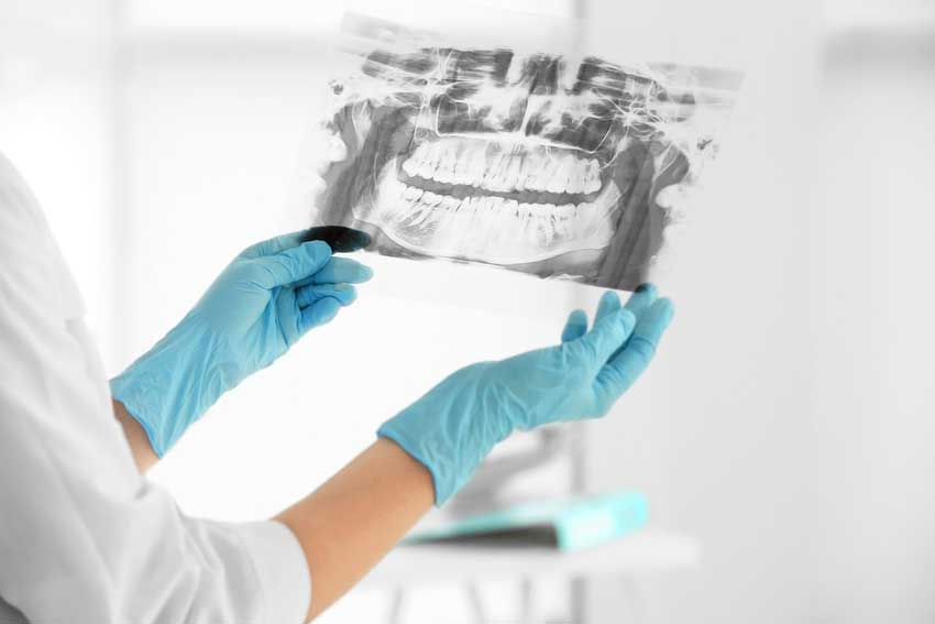
Dental x-rays are a common imaging tool used by dentists, and they are a crucial part of most people’s dental visits. They help a dentist to determine if there are any underlying problems with a patient’s teeth, that are not clinically visible to the naked eye. They enable the dentist to see what is going on in-between the teeth and under the gums. Dental x-rays are critical in identifying cavities, boneloss associated with gum disease, impacted teeth, as well as many other oral pathologies.
You may be worried about the use of x-rays within a dentist’s office, and what kind of risks come with having them done. If you’re curious about how dental x-rays work, and whether or not they’re really safe to use, these points should help enlighten you on the matter.
Why a Dental X-Ray is needed
Dental x-rays are typically performed once a year by a dentist, to ensure that their records on a patient’s oral health are up to date. They can also be carried out more often, if a dentist feels the need to track a patient’s dental problem or treatment at a given moment.
There are quite a few factors involved in how often dental x-rays will be carried out including a patient’s current oral health, their age,if they have a history of gum disease and if the patient has any dental concerns or continuing treatment.
Children may need more dental x-rays taken than adults, in order to monitorthe development of their teeth and jaw and also because they are more likely to be affected by tooth decay than adults.
The Types of Dental X-Rays
There are two main types of dental x-rays: intraoral and extraoral. Intraoral x-rays are taken with the film or the sensor inside the mouth and provide the dentist the detail needed to diagnose cavities, and check the health of the root and surrounding bone. Extraoral x-rays are taken with the film or sensor outside the mouth and are most often used to identify problems with the jaw and skull, as well as to look for impacted teeth. Let’s go through some of the most common x-rays taken at the dental office:
Bitewing – Used to check for cavities in between teeth by having the patient bite down on a special piece of paper. This x-ray also allows the dentist to see how the crowns of the teeth line up.
Panoramic – Used for a variety of issues, but most commonly used to check for wisdom teeth, or when evaluating a patient for dental implants. The x-ray machine will rotate around a patient’s head during this procedure.
Occlusal – Used to track the development and placement of an entire arch of teeth or section of teeth either in the upper or lower jaw
Periapical – Used to check for abnormalities in the root of the tooth and surrounding bone structures. This x-rays show the entire tooth from the crown to the root where the tooth attaches into the jaw.
The Risks of a Dental X-Rays
Most people are concerned about dental x-rays due to the fact that radiation is involved. However, the amount of radiation used during an x-ray is extremely small.
Many safeguards are taken during x-rays, to limit the amount of radiation that the patient receives. A‘bib’ made out of lead is placed over certain parts of a patient’s body, such as the pelvic region, or the chest and abdomen which prevents any excess radiation from penetrating the body, due to their vital organ placement.
Digital dental x-rays use an electronic sensor instead of the traditional x-ray film to capture images. These digital x-rays reduces radiation exposure to patients up to 90 percent compared to the already low exposure of traditional x-rays.
What Happens Next
Once the patient’s x-rays have been taken, the dentist will review them to check for problems in the teeth and gums and even the underlying bone structure of a person’s jaw.
If you are visiting a hygienist for a cleaning, he/she may use the x-rays to show you areas where you have tartar buildup or boneless so that you can be sure to brush and floss those areas better in the future. The dentist may go over any other findings from the x-rays after the cleaning is done. If there are any issues, with the use of x-rays the dentist will be able to detect the problems early in their development and treated.
The combination of a visual oral exam and x-rays provides the dentist with the necessary information to make sure that the patient can look forward to a future of continued or better oral health.

